
Associated Terms: Auricular Hematoma, Boxer Ear
Overview:
Aural hematoma is an abnormal collection of blood between the cartilage of the ear and the skin. It usually arises from a self-inflicted injury from scratching at inflamed/infected/painful ears and head shaking. Underlying causes include any condition that results in otitis externa (infection or inflammation, or both, of the external ear canal).
Hematoma formation has also been associated with increased capillary fragility (e.g., as seen with Cushing’s disease). Aural hematoma is the most common result of physical injury or trauma to the pinna (the “flap” of the ear). The condition is common in dogs with chronic otitis externa and less common in cats.
Sources of irritation to the ear linked to the development of aural hematoma include:
- inflammation
- immune-mediated disease
- allergies
- parasites
- foreign bodies
- trauma (bite wound or blunt trauma)
Many animals will have an associated infection. Recurrence of the condition is common even if the underlying condition is resolved.
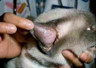
Swelling associated with an aural hematoma is most apparent on the concave inner surface of the pinna (Figure 1). The swelling is soft and warm in the early stages. With time, scar tissue will thicken and deform the ear, resulting in a cauliflower contracture.
Diagnostics:
A few simple tests may be performed to assess for an underlying cause of irritation/infection/pain.
- fine needle aspirate and cytology
- testing for underlying causes may include
- ear swabs – checking for bacterial, yeast, inflammatory cells or parasites
- endocrine testing
Pet owners should present their animals for examination and treatment early in the disease process before chronic changes occur to achieve the best results. Treatment options include needle aspiration and bandages, tube drainage systems and incisional drainage. The goals of surgery are to remove the hematoma, prevent recurrence and retain the natural appearance of the ears. Surgery typically includes making an incision on the underside of the ear flap to drain the fluid and is sometimes followed by placing several sutures to prevent fluid from building back up. A bandage is typically placed after surgery for a couple of days to help decrease swelling, discharge, and trauma from continued head shaking/scratching while any treatments for underlying causes are given time to reduce pain/inflammation/itching.
Aftercare and Outcome:
Deformity of the ear can occur even if the hematoma is drained. This typically will leave
- cosmetic alteration of the ear
- recurrence of the hematoma
- necrosis (death) of the pinna
A bandage or snood may be placed to protect the ear from infection and self-inflicted trauma. Infection can occur in the surgical site if the surgical wound is not managed appropriately by removing any wraps/bandages several times a day to all any moisture to dry, medication to be applied and blood supply remains unrestricted to the pinna if it is being held against the head with bandages/wraps.
Aural hematomas often reoccur even if they are properly treated initially and the underlying disease is appropriately managed, making this one of the most frustrating conditions for pet owners and pets!
Modified from: Auricular Hematoma, Boxer Ear. Accessed 7 JAN 25